Back Muscles Chart ~ Diagram Back Muscles Upper Back Human Anatomy Diagram Anatomy Human Body With Images Body Anatomy. Most of the time, back muscle pain is diagnosed then treated with little more than a prescription of rest, painkillers and muscle relaxants. Quadriceps (made of 4 muscles): Some of these muscles are quite large and cover broad areas. The deltoid, teres major, teres minor, infraspinatus, supraspinatus (not shown) and subscapularis muscles (not shown) all extend from the scapula to the humerus and act on the shoulder joint. The muscles of the back are a group of strong, paired muscles that lie on the posterior aspect of the trunk they provide movements of the spine, stability to the trunk, as well as the coordination between the movements of the limbs and the back muscles are divided into two large groups:
Your clients will thank you for it! It is attached to the calcaneus and is pulled by 3 flexor muscles: Most of the time, back muscle pain is diagnosed then treated with little more than a prescription of rest, painkillers and muscle relaxants. Some of these muscles are quite large and cover broad areas. An extremely strong tendon attached to the heel.
Muscle Diagram Of The Back Posterior Front Anterior from www.alpha-athlete.com October 28, 2020 reading time: Again, nerve damage associated with these symptoms can be permanent if not treated immediately. Molly smith dipcnm, mbant • reviewer: The muscles of the lower back help stabilize, rotate, flex, and extend the spinal column, which is a bony tower of 24 vertebrae that gives the body structure and houses the spinal cord.the spinal. The muscles of the back are a group of strong, paired muscles that lie on the posterior aspect of the trunk they provide movements of the spine, stability to the trunk, as well as the coordination between the movements of the limbs and the back muscles are divided into two large groups: Others, like sumo deadlifts, have been shown in emg studies—and in the trenches—to focus more on other muscle groups than the back. Nerves in your lower back. There are three different muscle groups found in the back:
Superficial muscles of the back are located directly deep towards the skin along with superficial fascia.they are occasionally called the appendicular group as these muscles are mainly associated with activities of the appendicular skeleton.
Quadriceps (made of 4 muscles): Muscles found in the superficial group include rhomboid major, rhomboid minor, levator scapulae, trapezius, latissimus dorsi. Related posts of muscles of the lower back and hip diagram muscle anatomy neck. Some of these muscles are quite large and cover broad areas. Function of the back muscles there are several individual muscles within the back anatomy, and it's important to take a quick look at all of Again, nerve damage associated with these symptoms can be permanent if not treated immediately. Muscle anatomy neck 12 photos of the muscle anatomy neck dog neck muscle anatomy, front neck muscle anatomy, muscle anatomy neck, muscle anatomy of neck and shoulder, neck muscle anatomy chart, human muscles, dog neck muscle anatomy, front neck muscle anatomy, muscle anatomy neck, muscle anatomy of neck and. Most of the time, back muscle pain is diagnosed then treated with little more than a prescription of rest, painkillers and muscle relaxants. Select a muscle group under each area to see the corresponding trigger points, referred pain patterns and stretches that should be performed along with pressure pointer treatment. Muscle anatomy diagram printable 12 photos of the muscle anatomy diagram printable muscle anatomy diagram printable, human muscles, muscle anatomy diagram printable Leaning back to straight vertical and all points in between. A strain can be an injury to a tendon attachment from muscle to bone. The extensor muscles are attached to back of the spine and enable standing and lifting objects.
We think this is the most useful anatomy picture that you need. Select a muscle group under each area to see the corresponding trigger points, referred pain patterns and stretches that should be performed along with pressure pointer treatment. Anatomynote.com found anatomy of back muscles diagram from plenty of anatomical pictures on the internet. These muscles include the large paired muscles in the lower back, called erector spinae, which help hold up the spine, and gluteal muscles. Enter the answer length or the answer pattern to get better results.
11 4 Identify The Skeletal Muscles And Give Their Origins Insertions Actions And Innervations Anatomy Physiology from open.oregonstate.education Chart of major posterior muscles. Deep back muscles diagram the superficial layer contains the splenius cervicis and splenius capitis muscles. Brings shoulders and arms back to body. Other muscles are small and cover much less space. Muscles found in the superficial group include rhomboid major, rhomboid minor, levator scapulae, trapezius, latissimus dorsi. The superior part of the appendicular skeleton that includes clavicle, scapula, and humerus, is attached to the axial skeleton that consists of skull. Back muscle anatomy chart 12 photos of the back muscle anatomy chart back muscle anatomy chart, lower back muscle anatomy chart, human muscles, back muscle anatomy chart, lower back muscle anatomy chart. A strain can be an injury to a tendon attachment from muscle to bone.
See back muscles and low back pain.
Five pairs of lumbar spinal nerves labeled l1 to l5 branch off your spinal cord and exit through small holes between the vertebrae. Some of these muscles are quite large and cover broad areas. Leaning back to straight vertical and all points in between. The muscles of the lower back help stabilize, rotate, flex, and extend the spinal column, which is a bony tower of 24 vertebrae that gives the body structure and houses the spinal cord.the spinal. No matter if you are a complete novice wanting to build muscle fast or an experienced muscle builder looking for that elusive muscle building routine that will promote new muscle growth. A strain can be an injury to a tendon attachment from muscle to bone. The superior part of the appendicular skeleton that includes clavicle, scapula, and humerus, is attached to the axial skeleton that consists of skull. When back development is the goal, stick to one of these variations. Symptoms of muscle pain include: Back muscle anatomy chart 12 photos of the back muscle anatomy chart back muscle anatomy chart, lower back muscle anatomy chart, human muscles, back muscle anatomy chart, lower back muscle anatomy chart. 1) make midline incision along spines of vertebrae 2) extend from This includes foot drop, a condition where the muscles of the leg and foot are too weak to raise the foot up as the individual attempts to walk. Anatomy chart courtesy of fcit the latissimus dorsi muscles (also known as the lats) are the largest muscles of the back.
The muscles of the lower back help stabilize, rotate, flex, and extend the spinal column, which is a bony tower of 24 vertebrae that gives the body structure and houses the spinal cord.the spinal. They extend and rotate the head and neck. Back pain symptoms chart view back pain chart pdf. The most common type of back pain is muscle pain—also called muscle strain or soft tissue strain. Anatomynote.com found anatomy of back muscles diagram from plenty of anatomical pictures on the internet.
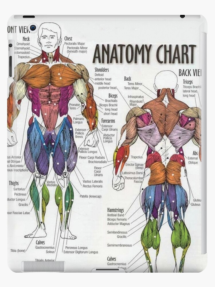
Anatomy Chart Muscle Diagram Ipad Case Skin By Superfitstuff Redbubble from ih1.redbubble.net When back development is the goal, stick to one of these variations. The extrinsic back muscles, which lie most superficially on the back. Back pain symptoms chart view back pain chart pdf. Five pairs of lumbar spinal nerves labeled l1 to l5 branch off your spinal cord and exit through small holes between the vertebrae. Superficial back muscles, intermediate back muscles and intrinsic back muscles.the intrinsic muscles are named as such because their embryological development begins in the back, oppose to the superficial and intermediate back muscles which develop elsewhere and are therefore classed as extrinsic muscles. The most common type of back pain is muscle pain—also called muscle strain or soft tissue strain. The deltoid, teres major, teres minor, infraspinatus, supraspinatus (not shown) and subscapularis muscles (not shown) all extend from the scapula to the humerus and act on the shoulder joint. Muscles found in the superficial group include rhomboid major, rhomboid minor, levator scapulae, trapezius, latissimus dorsi.
A strain can be an injury to a tendon attachment from muscle to bone.
When back development is the goal, stick to one of these variations. Superficial muscles of the back are located directly deep towards the skin along with superficial fascia.they are occasionally called the appendicular group as these muscles are mainly associated with activities of the appendicular skeleton. Function of the back muscles there are several individual muscles within the back anatomy, and it's important to take a quick look at all of Quadriceps (made of 4 muscles): Dimitrios mytilinaios md, phd last reviewed: Related posts of back muscles chart muscle anatomy diagram printable. Listed below are common areas of pain, or you can download a copy here. Other muscles are small and cover much less space. We think this is the most useful anatomy picture that you need. No matter if you are a complete novice wanting to build muscle fast or an experienced muscle builder looking for that elusive muscle building routine that will promote new muscle growth. Some of these muscles are quite large and cover broad areas. The muscle building industry is a mine field and i want to help people that are determined to build muscle naturally. Nerves in your lower back.